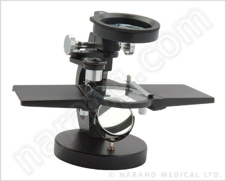
39 dissecting microscope diagram with labels
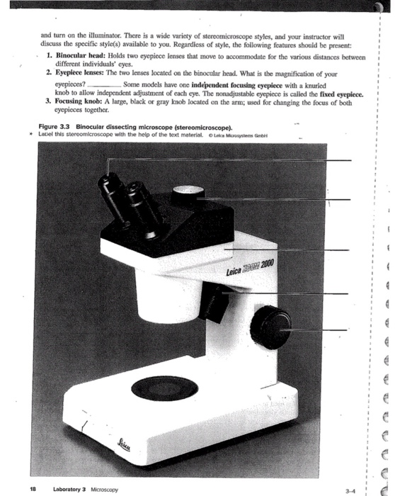
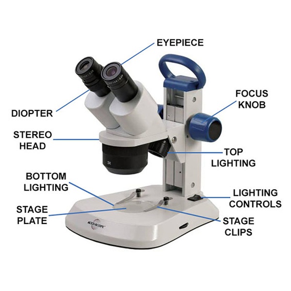
Parts of Stereo Microscope (Dissecting microscope) – labeled ... If you would like to learn optical components of a compound microscope, please visit Compound Microscope Parts – Labeled Diagram and their Functions, and this article. How to use a stereo (dissecting) microscope. Follow these steps to put your stereo microscopes in work: 1. Set your microscope on a tabletop or other flat sturdy surface where ... Conserved cell types with divergent features in human versus ... Aug 21, 2019 · After staining, sections were visualized on a fluorescence dissecting microscope (Leica) and cortical layers were individually microdissected using a needle blade micro-knife (Fine Science Tools ...
Ultra-high-throughput single-cell RNA sequencing and ... - Nature May 31, 2021 · Combining whole-transcriptome preindexing with standard droplet microfluidics, scifi-RNA-seq enables single-cell RNA-seq with massive throughput and built-in sample multiplexing.
Dissecting microscope diagram with labels
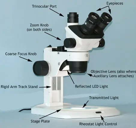
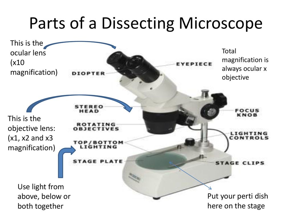
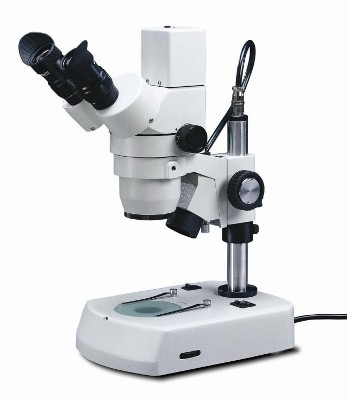
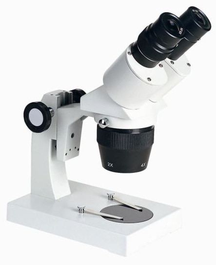
Shared and distinct transcriptomic cell types across ... - Nature Nov 01, 2018 · Single-cell transcriptomics of more than 20,000 cells from two functionally distinct areas of the mouse neocortex identifies 133 transcriptomic types, and provides a foundation for understanding ... Dissecting Stereo Microscope Parts and Functions Dissecting Stereo Microscope Parts and Functions Overview. Also known as a stereoscopic microscope, a dissecting microscope is a type of optical microscope commonly used for studying three-dimensional objects (3-D objects) as well as for dissecting biological specimen (e.g. insects and plant parts etc) at low magnification, between 2 and 100x depending on the microscope. COVID-19 immune features revealed by a large-scale ... - Cell Feb 03, 2021 · IGHV genes differentially used by moderate or severe COVID-19 patients compared with healthy controls and their intersections are shown with different colors. Venn diagram is used to show their overlaps with those published SARS-CoV-2 antibodies. Adjusted p values < 0.05 are indicated (two-sided unpaired Wilcoxon test).
Dissecting microscope diagram with labels. Botany Exam 1 Chs. 1, 2, 3, 4, 5, 6, 7, 12, and 16 Quizzes Simple, pinnately compound, and palmately compound leaves share some structures, but differ in others. To examine these similarities and differences in structure, click and drag the labels to their corresponding locations on the three diagrams. Note: Labels will be used more than once. COVID-19 immune features revealed by a large-scale ... - Cell Feb 03, 2021 · IGHV genes differentially used by moderate or severe COVID-19 patients compared with healthy controls and their intersections are shown with different colors. Venn diagram is used to show their overlaps with those published SARS-CoV-2 antibodies. Adjusted p values < 0.05 are indicated (two-sided unpaired Wilcoxon test). Dissecting Stereo Microscope Parts and Functions Dissecting Stereo Microscope Parts and Functions Overview. Also known as a stereoscopic microscope, a dissecting microscope is a type of optical microscope commonly used for studying three-dimensional objects (3-D objects) as well as for dissecting biological specimen (e.g. insects and plant parts etc) at low magnification, between 2 and 100x depending on the microscope. Shared and distinct transcriptomic cell types across ... - Nature Nov 01, 2018 · Single-cell transcriptomics of more than 20,000 cells from two functionally distinct areas of the mouse neocortex identifies 133 transcriptomic types, and provides a foundation for understanding ...
Stereo Microscope - Parts, Types and Uses - Laboratoryinfo.com
Dissecting microscope diagram - Lizzie Harper

20x-40x Industrial Binocular Stereo Microscope PCB Soldering Repairing Tool for Mobile Phone Clock Repairing and PCB Inspection

Multiple Choice Quiz on Compound Microscope Parts and Functions
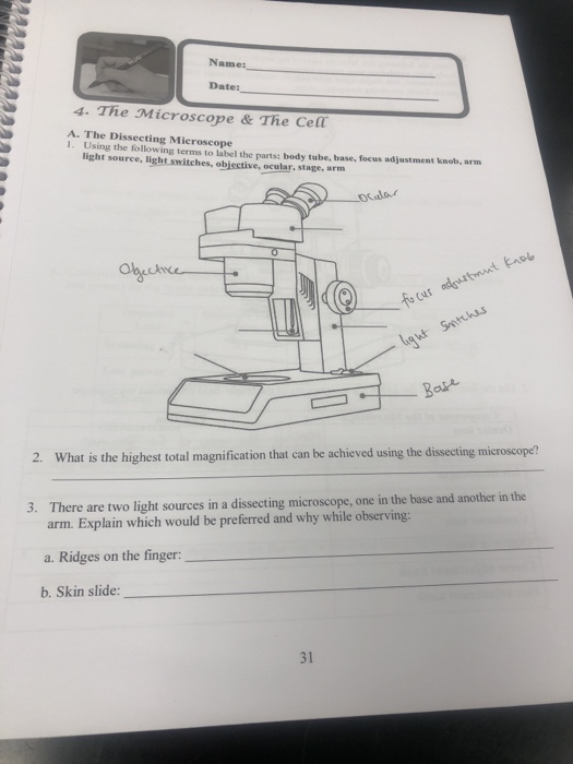
Name: Dates 4. The Microscope & The Cell A. The | Chegg.com

Types of Microscopes: Definition, Working Principle, Diagram ...
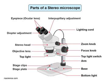
Parts of Stereo Microscope (Dissecting microscope) – labeled ...

Dissecting microscopes vs. Compound microscope
Dissecting Microscopes - ppt download

Microscope Parts & Functions - AmScope

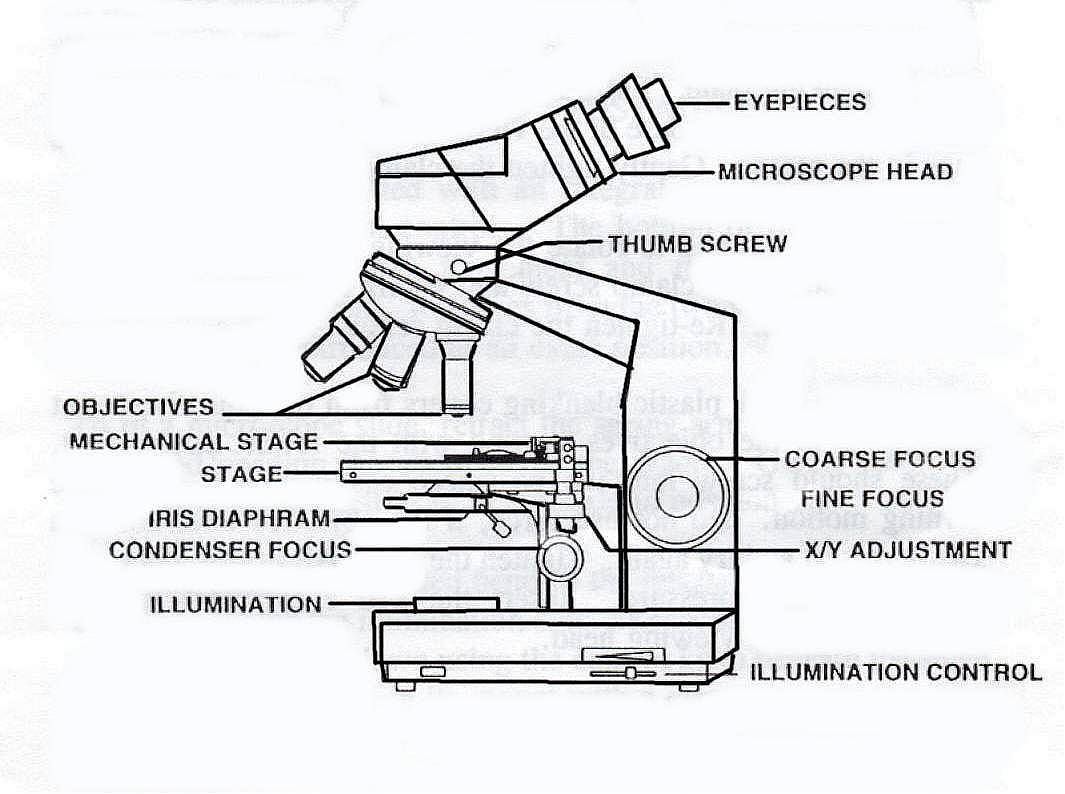
Compound Microscope Parts

Parts of Stereo Microscope (Dissecting microscope) – labeled ...
DC5-420TH Stereo Zoom Microscope from National Optical ...
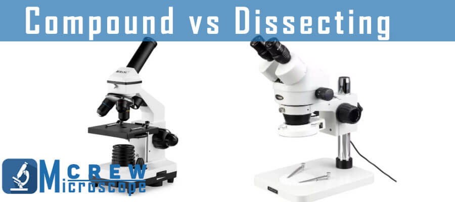
Difference between Compound and Dissecting Microscopes ...
Label the parts of the microscopes, and also answer | Chegg.com

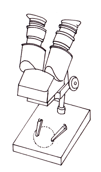
How to draw dissecting microscope step by step so easy

labeling dissecting microscope Diagram | Quizlet
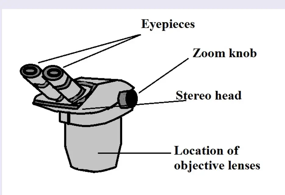
Dissecting Stereo Microscope Parts and Functions
PRACTICAL BOOKLET - BIOLOGY4ISC
1.2: Microscopes - Biology LibreTexts
Parts of Stereo Microscope (Dissecting microscope) – labeled ...

Tsetse biology, systematics and distribution, techniques

Digital Binocular Stereo Dissecting Microscopes with 2MP Camera and Software, WF10x and WF20x Eyepieces, 2X and 4X Objectives, 20X/40X/80X ...
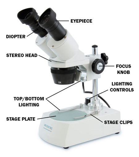
How to Use a Stereo Microscope and Science Lesson Ideas
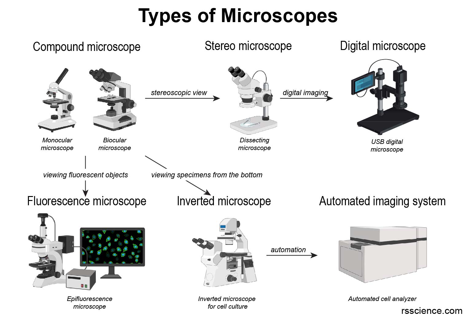
Different types of Microscopes – light microscope, electron ...

How To Draw A Microscope, Step by Step, Drawing Guide, by ...

Labelled Microscope with Functions | Microscope parts ...

dissecting microscope Diagram | Quizlet

Microscope With Labels clip art | Microscope parts, Science ...

02 Dissecting Microscope. A B Carrying a Microscope. - ppt ...
Dissecting Microscopes | Senior Dissecting Microscopes ...

How to Draw a Simple Microscope Diagram
Dissecting Microscope Uses - New York Microscope Company
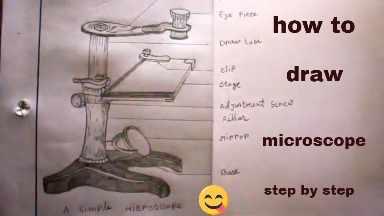
How TO Draw simple microscope step by step/simple microscope drawing/for science project
Simple Microscope - Parts, Functions, Diagram and Labelling ...
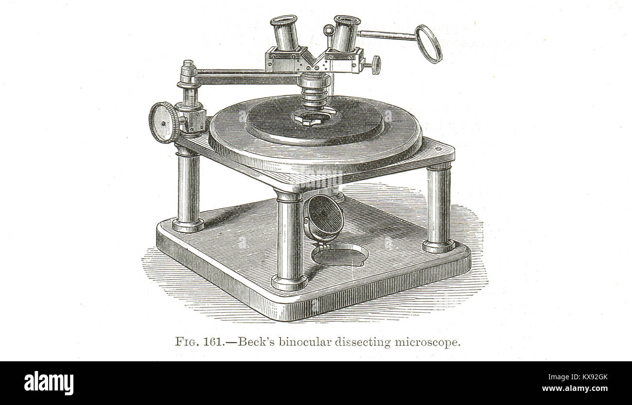
Microscopes. (a) Binocular dissecting microscope. (b ...

Tsetse biology, systematics and distribution, techniques

5 Important Types of Microscopes used in Biology (With Diagram)
Dissecting microscope hi-res stock photography and images - Alamy
Post a Comment for "39 dissecting microscope diagram with labels"